

Multi-analytical study of a Ptolemaic painted Egyptian textile cartonnage mask
Typological assessment and dating of the studied mask
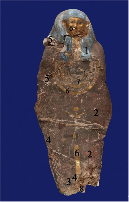
The studied object is a complete polychrome textile cartonnage mummy mask with a unified, non-segmented frontal configuration, comprising an integrated head and upper body with a symmetrically arranged tripartite wig. The gilded oval face exhibits idealized features, including almond-shaped eyes and proportionate facial elements, consistent with standardized funerary representation. The decorative scheme includes a broad-collar motif and a band of gilded hieroglyphic inscription, applied over a smooth preparatory layer. The continuity of the surface and the controlled execution of polychromy and gilding indicate a coherent and well-established workshop practice. These structural and stylistic features—particularly the absence of segmentation and the regularity of facial modeling—closely match established Ptolemaic typologies. Comparison with securely dated parallels (Fig. 2), including the Coffin of Pedi-Osiris, the Inner Coffin of Meret-it-is, and cartonnage from Akhmim, confirms strong correspondence in form and decorative conventions4,21. Accordingly, the mask can be confidently attributed to the Ptolemaic Period (2nd–1st centuries BCE) based on converging typological evidence 22.
A Coffin of Pedi-Osiris, Museum of Fine Arts, Houston; B Inner Coffin of Meret-it-is, Nelson-Atkins Museum of Art, Kansas City; C Ptolemaic cartonnage from Akhmim, British Museum. These parallels were used for stylistic dating of the studied mask.
Manufacture technique of the cartonnage (description of object)
Although the precise provenance and archeological context of object no. 1032 remain unknown; its construction was examined through direct observation and microstructural analysis. The internal stratigraphy and surface morphology of the cartonnage mask were investigated using digital optical microscopy and scanning electron microscopy (SEM) to document layer organization, material interfaces, and condition-related features (Fig. 3).

A Digital microscopy image showing the superimposed painted layer, preparatory ground layer, and underlying textile support; B SEM image of a detached fragment illustrating the multilayered cartonnage structure composed of linen support, ground layer, and paint layer.
Figure 3A shows a digital microscopy image of a polychrome area where three distinct zones are visible and marked numerically. The upper blue region (3) corresponds to the painted layer, which appears uneven and partially degraded, with areas of pigment loss and surface irregularity. Beneath it, the light-colored region (2) represents the preparatory ground layer, characterized by a compact and relatively smooth appearance. The lower brownish zone (1) corresponds to the underlying support, where a more irregular and coarse texture is visible, likely associated with the textile substrate and degraded material. The boundary between these zones is clearly distinguishable, demonstrating the superposition of the painted layer over a preparatory ground applied onto the support.
Figure 3B presents an SEM image of a detached fragment from the edge of the cartonnage, clearly illustrating the multilayered structure. The lower portion of the image, composed of elongated, interwoven fibrous elements, corresponds to the linen textile support. Above this, a dense and compact layer with a relatively smooth surface represents the preparatory ground layer. This layer appears continuous and well-adhered to the textile fibers. The uppermost surface, where preserved, corresponds to the pigmented layer, which shows cracking and partial detachment, visible as irregular fractures across the surface. The observed structure confirms the typical cartonnage manufacturing technique, consisting of a textile base coated with a mineral preparatory layer to provide a suitable surface for polychrome decoration.
Identification of textile support
The textile forming the structural base of the cartonnage was examined using digital optical microscopy, SEM, and XRD to identify its fiber type, manufacturing features, and general state of preservation.
Digital optical microscopy (Fig. 4A–C) reveals a plain 1/1 weave composed of cellulosic fibers exhibiting longitudinal striations and ribbon-like flattening, features consistent with linen-type textiles widely used in ancient Egypt. Although optical microscopy alone cannot confirm botanical origin, the observed morphology supports this attribution. The images also show fiber darkening, particulate contamination, and mechanical damage, including fraying, tearing, and surface scratches, indicative of long-term burial and environmental exposure.

A Darkened fibers showing oxidative aging and burial alteration; B dust and soil particles trapped between fibers; C mechanical damage including fraying, tearing, scratches, and fiber loss.
In Fig. 4A, pronounced fiber darkening is associated with oxidative aging, burial-related deposits, and degradation products, accompanied by reduced reflectivity and increased brittleness, reflecting chemical and structural weakening of cellulose23. Figure 4B shows dense accumulations of dust and soil particles entrapped between fibers, contributing to abrasion, stiffening, and reduced flexibility. Mechanical deterioration is evident in Fig. 4C through fiber breakage, fraying, and tearing, resulting from environmental fluctuations and inherent fragility.
SEM analysis was conducted to further characterize the microstructure of the textile support and assess fiber-level deterioration resulting from aging, burial conditions, and long-term environmental exposure. SEM imaging provided high-resolution visualization of the weave organization, fiber surface morphology, and micro-damage features not discernible by optical microscopy.
SEM analysis (Fig. 5A–E) provides higher-resolution characterization of the textile microstructure. The images confirm a plain 1/1 weave with fibrillar morphology typical of plant-based fibers, consistent with linen used in Egyptian cartonnage24. Degradation features include transverse cracking (Fig. 5C), indicating repeated shrinkage–swelling cycles; longitudinal splitting (Fig. 5D), reflecting hydrolytic degradation; and surface abrasion with adherent particles (Fig. 5E), linked to burial contamination and environmental deposits25. Additional features such as micro-scratches, shallow slits, partial delamination, and micro-cavities indicate advanced mechanical deterioration, loss of amorphous cellulose regions, and possible microbial activity26. Despite these effects, the overall weave structure remains preserved

A, B Plain 1/1 weave structure of the linen support and fiber arrangement; C transverse cracking; D longitudinal splitting; E surface abrasion, scratches, and adhering particles caused by deterioration.
XRD analysis (Fig. 6) provides complementary molecular-level insights, showing reduced crystallinity at characteristic cellulose Iβ peaks (14.9°, 22.7°, 34.5° 2θ) and the presence of an amorphous halo (18–25° 2θ). These features indicate glycosidic bond scission and oxidative and hydrolytic breakdown of cellulose, highlighting the structural compromise of the fibers and the need for careful conservation handling19,27.
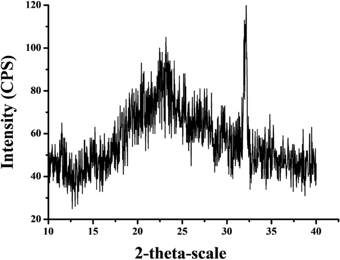
X-ray diffraction pattern showing cellulose-related peaks and reduced crystallinity associated with degradation of the linen fibers.
Identification of the ground layer
The preparatory ground layer (gesso) forms the intermediate interface between the linen textile support and the overlying polychrome decoration. Its structure, composition, and state of preservation were examined through complementary microscopic and elemental analyses on both surface (Fig. 7).

A, B Digital microscopy of exposed white ground layer and interface with textile support; C SEM image showing compact crystalline matrix; D EDS spectrum confirming calcium-rich composition; E portable XRF spectrum supporting calcite-rich ground material.
Digital microscopy of partially exposed areas (Fig. 7A) reveals a compact, fine-textured white layer with localized micro-cracking at the interface with the overlying pigmented layers. Minor pigment flaking is observed in degraded zones; however, the underlying ground layer remains largely continuous. Higher-magnification images (Fig. 7B) show a well-defined and adherent transition between the linen textile support and the ground layer, with limited evidence of lifting or separation at the interface.
SEM observations (Fig. 7C) provide detailed insight into the microstructure of the ground layer. The layer appears as a dense and continuous matrix composed of fine angular to sub-angular crystalline particles. These particles are visible in the image as bright, closely packed grains that are evenly distributed across the surface. In contrast, the darker, elongated features correspond to the underlying linen fibers, which are partially embedded within the ground layer. The intimate contact between the crystalline matrix and the textile substrate indicates strong adhesion and effective penetration of the preparatory material into the fibrous structure. Furthermore, the absence of significant voids, extensive cracking, or interfacial separation suggests that the ground layer is well preserved and maintains good mechanical stability 28.
EDS analysis of the ground layer (Fig. 7D and Table S1) shows a dominant presence of calcium (Ca), consistent with a calcium carbonate composition corresponding to calcite. Minor sulfur (S) signals indicate the presence of small amounts of gypsum (CaSO4·2H2O), while occasional silicon (Si) peaks suggest trace siliceous inclusions, likely related to quartz impurities. The elemental composition and particle morphology are consistent with carbonate-based gesso layers commonly reported in Ptolemaic cartonnage production9,10. SEM observations further indicate a compact and cohesive microstructure, with no pronounced porosity, cracking, or interlayer delamination.
In situ pXRF analysis (Fig. 7E and Table S2) corroborates the EDS results, showing calcium as the dominant elemental component, accompanied by trace sulfur and very low concentrations of iron, copper, strontium, and lead. The limited presence of heavy elements and silicate-related signals supports the use of a calcite-rich ground material with a relatively high degree of purity.
Overall, the ground layer is characterized by a uniform thickness, compact microstructure, and close adhesion to the textile support, forming a stable preparatory layer for the application of polychrome decoration and gilding.
Investigation of blue pigment
The blue pigment applied to the tripartite wig and decorative bands was examined to assess its composition, microstructure, and state of preservation (Fig. 8).

A–F Digital microscopy images showing preserved and deteriorated blue-painted areas; G SEM image of pigment particles embedded in the ground layer; H EDS spectrum identifying Egyptian blue; I portable XRF spectrum confirming calcium–copper silicate composition.
Digital microscopy observations (Fig. 8A–F) reveal variable preservation states across the blue-painted areas. Well-preserved regions (Fig. 8A) display a fine crystalline texture, a vivid turquoise-blue hue, and strong adhesion to the underlying ground layer. Moderately altered areas (Fig. 8B–C) show micro-cracking, pigment granulation, and slight thinning at the edges of the painted zones. In more severely deteriorated regions (Fig. 8D–F), the pigment surface exhibits increased roughness, fissuring, partial discoloration toward grayish or darkened tones, pigment detachment, and accumulation of fine debris. These features indicate localized degradation of the pigment–binder system and interaction with environmental agents.
SEM imaging (Fig. 8G) reveals angular to platy pigment particles embedded within the upper portion of the preparatory ground layer. The pigment grains appear well integrated within the matrix, although localized micro-cracks are observed traversing the ground and pigment interface. Evidence of partial binder loss is reflected in the presence of small voids and reduced cohesion between adjacent particles. In addition, thin surface films are detected on some pigment grains, likely corresponding to secondary phases enriched in calcium and sulfur. These microstructural alterations are consistent with the surface dulling, cracking, and textural irregularities observed under digital microscopy, confirming ongoing physicochemical degradation processes affecting the pigment layer.
EDS analysis (Fig. 8H and Table S3) reveals dominant Si (39.99 wt%, 53.97 at%), Cu (39.76 wt%, 23.72 at%), and Ca (13.36 wt%, 12.64 at%), along with minor Al (6.88 wt%, 9.67 at%). The combined presence of Si and Cu, together with Ca, is consistent with a calcium copper silicate phase, characteristic of cuprorivaite (CaCuSi4O10) and supports identification of the pigment as Egyptian blue6,8. Although Ca could partially originate from the underlying ground layer, particularly given the stratigraphic proximity, SEM observations (Fig. 8G) demonstrate that these elements are spatially associated within the pigment particles themselves. This microstructural association supports their incorporation within a single mineral phase rather than representing a signal exclusively derived from the substrate. The relative elemental proportions—especially the high Si content coupled with substantial Cu and Ca—further reinforce the identification of the pigment as Egyptian blue.
pXRF analysis (Fig. 8I and Table S4) corroborates the EDS results, revealing strong Ca (≈54.71%) and Cu (≈28.68%) signals, along with detectable Si. Although the elevated Ca content may be partially influenced by the underlying preparatory layer due to the greater penetration depth of pXRF, its consistent association with Cu and Si—supported by SEM–EDS microstructural observations—indicates the presence of a calcium copper silicate phase.
This elemental combination clearly distinguishes the pigment from copper carbonate blues such as azurite (Cu3 (CO3)2(OH)2), which lack a silicate component and would exhibit a markedly different elemental signature. Localized enrichment in chlorine (Cl ≈ 14.81%) is observed in deteriorated areas, suggesting salt-related alteration processes 29.
Collectively, these results confirm the use of Egyptian blue30 and highlight microstructural evidence of ongoing alteration, including cracking, binder loss, and surface modification by secondary phases 31.
Investigation of red pigment
The red pigment used in the decorative elements of the cartonnage mask was systematically evaluated to elucidate its microstructural organization, compositional profile, and extent of degradation (Fig. 9).

A–D Digital microscopy images showing varying preservation states of red-painted zones; E SEM image of compact pigment grains over the ground layer; F EDS spectrum identifying vermilion; G portable XRF spectrum showing mercury with associated iron-rich components.
Digital microscopy observations (Fig. 9A–D) reveal variable preservation states across the red-painted areas. Well-preserved regions (Fig. 9A) display a dense, matte red surface with no visible micro-cracking, indicating good cohesion between the pigment layer and the underlying ground. Moderately altered zones (Fig. 9B) exhibit slight chromatic shifts toward orange-brown tones, fine fissures, and embedded particulate matter. In more deteriorated areas (Fig. 9C), the pigment layer appears thinned and discontinuous, with partial exposure of the white preparatory ground and loosely packed pigment particles. The most severely affected regions (Fig. 9D), particularly near folds and structural transitions, show pronounced cracking, flaking, surface soiling, and localized delamination.
SEM imaging (Fig. 9E) shows angular to sub-rounded pigment grains ranging from approximately 3 to 10 µm in size, forming a compact layer directly over the calcite- and gypsum-rich preparatory ground. No distinct intermediate layer is observed between pigment and ground. The pigment particles are embedded within a partially degraded organic matrix, with micro-voids and localized porosity that correspond to the deterioration features observed under optical microscopy.
EDS spectra (Fig. 9F and Table S5) reveal a dominant mercury (Hg) signal (71.58 wt%), accompanied by sulfur (S: 4.04 wt%), confirming the presence of mercury sulfide (HgS), consistent with vermilion10. No iron (Fe) signal is detected in the analyzed EDS spot, indicating that the examined micro-area is predominantly composed of a relatively pure HgS phase. In contrast, pXRF analysis (Fig. 9G and Table S6) identifies Hg (28.93%) together with a significant Fe content (14.75%), as well as minor Ti, K, and Sr. The discrepancy between EDS and pXRF results reflects differences in analytical scale and sampling volume: whereas EDS probes a highly localized microscale region, pXRF analyzes a broader area that may include adjacent layers and compositional heterogeneity, The detected Fe is therefore attributed to iron-rich phases, most likely hematite, either present as an underlying or adjacent red layer or as part of a mixed pigment application, which may also explain tonal variations observed in the painted surface12. The absence of arsenic (As) excludes arsenic sulfide pigments such as realgar (As4S4), while the lack of lead (Pb) rules out red lead (Pb3O4). No elemental signatures indicative of modern synthetic red pigments were detected. Elevated chlorine levels (Cl ≈ 41.86%) observed in deteriorated areas are attributed to salt contamination and post-depositional alteration processes rather than the original pigment composition. These results confirm that vermilion was intentionally employed for its vivid red hue. The presence of iron detected by pXRF is interpreted as evidence of hematite, likely associated with pigment admixture, stratigraphic overlap, or variability in raw material sources, which may also account for tonal differences across the painted surface. Such practices are consistent with pigment preparation and application techniques documented for the Ptolemaic period.
Although vermilion is generally considered a stable pigment, the observed flaking, discoloration, and weakening of the paint layer are attributed to binder degradation, mechanical stress, and instability of the preparatory ground (gesso). The detection of chloride salts suggests a potential risk for HgS alteration and darkening; however, no evidence of transformation to elemental mercury was identified32.
Identification of black pigment
The black pigment employed in the cartonnage mask was systematically examined to elucidate its microstructural features, compositional characteristics, and degree of preservation (Fig. 10).

A–C Digital microscopy images of preserved and deteriorated black-painted areas; D SEM image of dense pigment layer; E EDS spectrum indicating iron oxide black pigment; F portable XRF spectrum confirming iron-rich composition.
Digital microscopy observations reveal variable preservation states of the black-painted areas. Well-preserved regions (Fig. 10A) display a uniform, deep black tone with a fine granular texture and strong adhesion to the underlying white ground, with no visible cracking or fading. Moderately altered zones (Fig. 10B) show slight graying, reduced color saturation, and fine surface fissures, accompanied by minor surface abrasion and particulate contamination. In more severely degraded areas (Fig. 10C), the pigment layer exhibits pronounced fading, brittleness, and a chalky appearance, with extensive cracking, thinning, and localized losses that expose the underlying gesso or textile support.
SEM imaging (Fig. 10D) reveals a thin, compact pigment layer applied directly over the white preparatory ground. The black pigment appears as a dense, fine-grained matrix lacking visible crystalline features at the examined magnifications. The pigment particles are irregular in shape, ranging from approximately 0.5 to 3 µm in size, and form a continuous layer with no discernible intermediate strata between pigment and ground 1.
EDS spectra (Fig. 10E and Table S7) reveal a simple but highly diagnostic elemental profile dominated by Fe (70.05 wt%) and O (29.95 wt%), yielding an Fe:O atomic ratio consistent with magnetite (Fe2O4). The strong, homogeneous Fe signal and the absence of significant C indicate a mineral black pigment rather than carbonaceous materials such as soot, lampblack, or bone black, which would show overwhelming carbon enrichment. Minor traces of Ca and Si observed in some points likely derive from the underlying calcite-based ground layer or incidental sediment inclusions rather than the pigment itself.
Portable XRF measurements (Fig. 10F and Table S8) corroborate this interpretation: Fe is the dominant chromophore (27.59%), and no elements characteristic of alternative historical black pigments—such as Mn (for manganese black) or Pb (for lead-black mixtures)—were detected. Elevated Cl (36.7%) and K (21.78%) are attributed to burial-related salt contamination rather than original pigment composition, a common occurrence in archeological cartonnage. The minor Ti and Cu signals represent natural accessory minerals within the iron oxide source or trace environmental accretions.
Together, the EDS and pXRF datasets provide consistent evidence that the black pigment used on the mask is a natural iron-oxide black, specifically magnetite. This material was widely employed in Ptolemaic and Roman workshops for outlines, detailing, and inscriptions due to its opacity, stability, and ready availability from iron-rich geological deposits12,21. The mineralogical purity and uniform application observed here reflect a deliberately selected, high-quality black pigment rather than a carbon-based or composite mixture.
Investigation of green pigment
The green pigment decorating the cartonnage mummy mask was comprehensively evaluated to define its microstructural organization, elemental composition, and extent of degradation (Fig. 11).

A–C Digital microscopy images showing preserved and altered green-painted areas; D SEM image of pigment particles and binder matrix; E EDS spectrum identifying malachite; F portable XRF spectrum confirming copper-based green pigment.
Digital microscopy observations reveal variable preservation states of the green-painted areas. Well-preserved regions (Fig. 11A) show a compact, finely textured green layer with uniform coloration and strong adhesion to the underlying white ground. Moderately deteriorated zones (Fig. 11B) exhibit uneven chromatic tones, fine fissures, micro-abrasions, and embedded particulate matter, indicating partial weakening of the pigment–binder–substrate system. Severely degraded areas (Fig. 11C) display powdering, discoloration, cracking, and localized detachment of the pigment layer, exposing the preparatory ground in some locations.
SEM imaging (Fig. 11D) reveals that the green pigment was applied directly over the white gesso layer. The pigment particles are angular to sub-round in morphology, typically ranging from approximately 3 to 12 µm in size, and are embedded within a partially degraded organic matrix. Micro-cracks, increased porosity, and localized separation at the pigment–ground interface are evident, particularly in areas corresponding to advanced surface deterioration observed under optical microscopy.
EDS analysis (Fig. 11E and Table S9) shows a compositional profile dominated by copper (Cu), accompanied by carbon (C) and oxygen (O), consistent with the basic copper carbonate mineral malachite (Cu2CO3(OH)2). Minor chlorine (Cl) is also detected, indicating localized alteration of the copper carbonate pigment to copper chloride phases under burial or post-depositional environmental conditions. Trace amounts of calcium (Ca) and sulfur (S) are attributed to contributions from the underlying calcite–gypsum gesso or to secondary sulfate contamination, in agreement with previously reported studies on Egyptian polychrome materials12.
In situ pXRF analysis (Fig. 11F and Table S10) corroborates the EDS results, showing clear detection of copper together with chlorine and calcium. Although calcium registers as a dominant element due to matrix effects and the measurement depth encompassing the preparatory ground, the presence of Cu confirms the copper-based nature of the green pigment. Trace detections of iron (Fe), strontium (Sr), and arsenic (As) are attributed to mineral impurities or environmental contamination. The elemental pattern excludes chromium-based green pigments such as viridian (Cr2O3·2H2O) and arsenic-containing copper greens such as emerald green(Cu(C2H3O2)2·3Cu(AsO2)2), neither of which exhibits the Cu–C–O association or copper–chloride signature observed here33.
Overall, the combined digital microscopy, SEM–EDS, and pXRF analyses identify malachite as the primary green pigment used on the mask. The pigment layer shows varying degrees of preservation, ranging from compact, well-adhered coatings to areas affected by cracking, powdering, and partial detachment, particularly in zones influenced by environmental stress and salt-related alteration31,34.
Identification of white pigment
The white pigment decorating the cartonnage mummy mask was comprehensively examined to define its microstructural organization, elemental composition, and degree of alteration (Fig. 12).

A–C Digital microscopy images showing preserved, cracked, and deteriorated white-painted areas; D SEM image of compact white layer over ground; E EDS spectrum identifying calcite-rich composition; F portable XRF spectrum confirming calcium-based white pigment.
Digital microscopy reveals three distinct preservation states. Well-preserved areas (Fig. 12A) exhibit a smooth, homogeneous white surface firmly adhered to the underlying ground, with no visible cracking, powdering, or particle separation. Moderately deteriorated zones (Fig. 12B) show fine micro-cracks, a slightly roughened surface texture, minor chromatic alteration toward yellowish or gray tones, and early-stage pigment loss, indicative of partial binder degradation associated with environmental cycling. Severely degraded regions (Fig. 12C) display chalking, fracturing, pigment detachment, and the incorporation of dust or fibrous inclusions, reflecting mechanical stress and prolonged exposure to moisture or salt-related processes.
SEM imaging (Fig. 12D) shows a thin, compact white pigment layer applied over a thicker preparatory ground composed of calcite and gypsum. The pigment layer consists of tightly packed fine grains with minimal internal porosity, indicating deliberate surface preparation and controlled application.
EDS spectra (Fig. 12E and Table S11) are dominated by calcium (Ca) and oxygen (O), consistent with the presence of calcium carbonate (calcite, CaCO3). Minor sulfur (S) is detected within 4·2H2O) or secondary sulfate contamination related to burial or post-depositional environmental exposure. Importantly, no lead (Pb) or zinc (Zn) signals are present, effectively excluding the use of white lead (2PbCO3·Pb(OH)2) or zinc white (ZnO), pigments that belong to later historical periods and were not available in ancient Egyptian painting practice 35.
The pXRF results (Fig. 12F and Table S12) corroborate the EDS data, confirming calcium as the dominant element with trace sulfur. The absence of significant heavy-metal elements further supports the identification of a calcium-based white pigment. The combined analytical evidence indicates that the white layer is composed predominantly of natural calcite, with minor gypsum either intentionally incorporated or introduced through post-depositional processes. Such calcium-based white materials are characteristic of traditional Egyptian gesso and white paint preparations12.
Overall, the analytical data confirm that the white pigment is primarily calcium carbonate, occasionally associated with gypsum, applied as a thin layer over a similar preparatory ground. Variations in preservation, including micro-cracking, surface soiling, and localized flaking, correlate with binder degradation and environmental stress rather than intrinsic pigment instability31,34.
Identification of gilded layers
The gilded surfaces of the cartonnage mummy mask were comprehensively examined to define their material composition, layer structure, and degree of alteration, especially in the facial region and ornamental details (Fig. 13).

A–C Digital microscopy images showing preserved, cracked, and detached gold leaf surfaces; D portable XRF spectrum confirming high-purity gold leaf applied over a calcium-rich ground layer.
Digital microscopy reveals three distinct preservation states. Well-preserved regions (Fig. 13A) exhibit a smooth, continuous, and highly reflective gold surface firmly adhered to the underlying preparatory layer, with only minor abrasions or isolated micro-cracks. Moderately deteriorated areas (Fig. 13B) show localized dulling, fine cracking, partial delamination, and edge curling of the gold leaf, consistent with adhesive weakening, mechanical stress, and humidity-related dimensional changes. Severely degraded zones (Fig. 13C) are characterized by extensive fragmentation, loss of gold leaf, and exposure of the underlying white ground or adjacent pigment layers, frequently accompanied by embedded dust particles and salt residues, indicating advanced deterioration.
The pXRF analysis (Fig. 13D and Table S13) shows a dominant gold (Au) signal, with intense Lα and Lβ emission lines confirming the presence of gold leaf. No detectable signals of copper (Cu), silver (Ag), lead (Pb), or chlorine (Cl) were observed, excluding the use of gold alloys, composite metallic foils, or corrosion-related alteration products. The absence of these elements indicates the application of high-purity, unalloyed gold. Calcium (Ca) and sulfur (S) peaks are also present and are attributed to the underlying preparatory ground, likely composed of gypsum (CaSO4·2H2O) and/or calcite (CaCO3). The relatively elevated Ca signal reflects the penetration depth of the pXRF beam, which partially samples the gesso layer beneath the extremely thin gold leaf. Minor signals of iron (Fe), potassium (K), and chromium (Cr) are interpreted as environmental particulates or post-depositional surface contamination rather than intentional constituents of the gilding layer. Overall, the analytical results confirm that the gilded areas were produced using thin, high-purity gold leaf applied over a calcium-rich ground layer. This material configuration is consistent with documented gilding practices employed in Ptolemaic and Roman-period cartonnage production28.
Identification of binding media
The binding media in ancient Egyptian polychrome cartonnage were essential for the cohesion of pigments, ground layers, and textile supports. These organic compounds functioned as both adhesives and pigment carriers, making their identification vital for understanding manufacturing techniques, assessing material stability, and guiding conservation. In this study, the binding media within the cartonnage mask stratigraphy were analyzed using FTIR-ATR, with spectra collected from the textile support and the painted/gilded decorative layers (Fig. 14).

A FTIR spectrum of adhesive residues from the textile support indicating proteinaceous animal glue; B FTIR spectrum from painted and gilded layers indicating polysaccharide binder consistent with gum Arabic.
Figure 14A shows the FTIR spectrum obtained from a sample collected from the textile support layer, where remnants of the original adhesive were still present. The spectrum exhibits absorption bands characteristic of proteinaceous materials, consistent with an animal-derived binder. Notable features include Amide I (∼1640 cm−1) and Amide II (∼1535 cm−1) bands, corresponding to C = O stretching and N–H bending vibrations, respectively, as well as a broad O–H/N–H stretching band around 3300–3400 cm−1and CH₂ bending at 1450 cm−1. These results indicate the presence of a proteinaceous adhesive of animal origin, as a primary adhesive between the layers of textile support and also the preparatory ground layer36.
Figure 14B displays the FTIR spectrum from samples taken from pigmented zones. In contrast to the textile layer, these spectra are dominated by absorption bands attributable to polysaccharide-based binders, primarily gum Arabic. Key features include a strong O–H stretching band around 3300 cm−1, C–H stretching at 2920 cm−1, and intense C–O–C and C–O stretching vibrations between 1030 and 1060 cm−1, typical of complex carbohydrates37. A weak carbonyl peak near 1730 cm−1 suggests minor oxidative aging of the gum. The absence of Amide I and II bands indicates that no proteinaceous materials were used in these decorative surface layers.
These results indicate a layer-specific use of binders: a proteinaceous animal-derived adhesive for the structural layers and gum Arabic for pigments, reflecting the technical knowledge of Ptolemaic artisans. From a conservation perspective, both materials are hygroscopic and sensitive to fluctuating humidity13,21.
Credit: Source link